
Dental Technology in Pottsville, PA
A clearer picture of what’s going on in your mouth, shown on a screen right next to you. Fellows & Smink Dental Associates uses digital imaging tools that help us catch problems early and explain them in language that makes sense.
What We Mean by Dental Technology in Pottsville PA
When patients ask about dental technology in Pottsville PA, they usually want to know two things. What are you going to use on me, and is it going to help me understand what’s happening? Both fair questions. We try to answer them before we pick up an instrument.
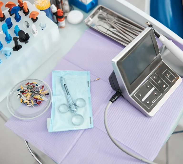
At Fellows & Smink Dental Associates, our diagnostic tools are built around digital X-rays read with the Overjet Iris system and small intraoral cameras that show the inside of your mouth on a screen. None of it is there for show. It’s there so Dr. Scott Fellows can find issues earlier, explain them clearly, and give you a real say in what comes next.
We’re a family practice on West End Avenue, and most of our patients come from Pottsville, Saint Clair, Minersville, Schuylkill Haven, and the surrounding Schuylkill County towns. The technology we use should make a visit feel more straightforward, not more complicated. That’s the bar.
How Digital X-Rays and the Overjet Iris System Work
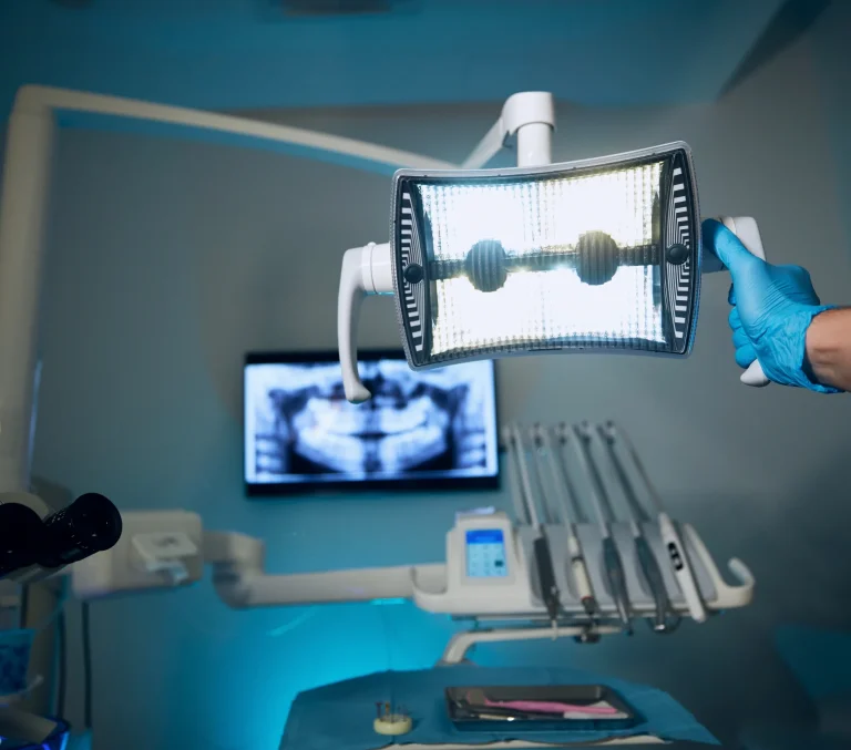
Digital X-rays use a small sensor instead of film. The image shows up on the screen in seconds, the radiation dose is significantly lower than the older film method, and we can zoom in on a specific tooth without retaking anything. For kids who don’t love sitting still and for older patients who want to be in and out, that speed matters.
The Overjet Iris system is artificial intelligence that reads X-rays alongside Dr. Fellows. It outlines areas of possible decay, measures bone levels around teeth, and flags things that are easy to miss with the eye alone. Dr. Fellows still makes the call on every diagnosis. The software is a second set of eyes, not a replacement for his.
What this means for you in the chair: when we say there’s a small cavity forming between two back teeth, you can see the outline on the same screen we’re looking at. No guessing. No taking our word for it. If something can wait and be watched, we’ll tell you that too.
What Intraoral Cameras Show You During an Exam
The intraoral camera is a small wand, about the size of a pen, with a light and a lens on the end. We move it through your mouth and the picture shows up live on the monitor. A cracked filling, a chipped edge, a piece of a tooth that’s wearing down faster than the rest, you’ll see it the same way we do.
This is especially useful when we’re talking about restorative work like crowns and bridges or a dental filling that’s starting to fail. Patients often say the same thing when they see the picture: oh, that’s what you mean. That moment of recognition makes the conversation about treatment a lot easier.
It also helps with prevention. If we can show a parent why their child’s back molar would benefit from dental sealants, or show an adult patient the gum tissue that’s starting to pull away from a tooth, the why becomes obvious. Pictures do more work than a paragraph of explanation.
What's Included When You Come in for Imaging
For most new patients, a first visit includes a set of digital X-rays, a full exam with Dr. Fellows, and intraoral photos of any areas worth a closer look. We walk through what’s on the screen with you. If there’s nothing to worry about, we say so. If there’s something to address, we map out the order it should happen in and what your insurance will likely cover.
For returning patients, we take updated X-rays at the intervals recommended for your age and risk level, usually less often than people expect. Bitewing X-rays for checking between teeth tend to be every one to two years for most adults. We don’t take images we don’t need.
Because we keep a full range of care under one roof, the same images carry through if you end up needing work in another department: a tooth extraction, a dental implant consultation, periodontal maintenance, or a referral to Dr. Fellows for restorative planning. You won’t be asked to repeat X-rays at a separate office across town. Same building, same records, same team.
How Imaging Is Billed Through Your PPO Plan
Diagnostic X-rays and exams are usually covered by PPO dental insurance at a high percentage, often at or near 100% for the preventive portion of your plan. We accept all PPO dental plans and file the claims for you, so you don’t have to chase paperwork after the visit.
If you don’t have coverage, we offer flexible payment options and will sit down to map out what makes sense for your month. For a closer look at how billing works at our office, you can read more on our dental insurance page or ask about payment plans when you call.
Why Better Imaging Changes the Visit
Catching a problem on an X-ray when it’s the size of a pinhead is a different conversation than catching it when it’s already a toothache. The first is a small filling on a Tuesday morning. The second can mean root canal therapy or an extraction and a longer road back. Better imaging shifts more of your care toward the first kind of visit.
It also keeps the visit honest. When you can see what we see, there’s no need to oversell anything. We tell you what’s urgent, what can wait, and what we’re going to keep an eye on at your next cleaning. That’s the way we’d want our own family treated, and it’s how we run the practice for yours.
If you’d like to meet the team and the tools in person, you can learn more about Dr. Scott Fellows or contact our Pottsville office to set up a first visit.
See the Technology for Yourself
Schedule a visit at our West End Avenue office and we’ll show you your X-rays and intraoral images on screen, then explain what they mean in plain language.